
Em-Lux benchtop imaging system
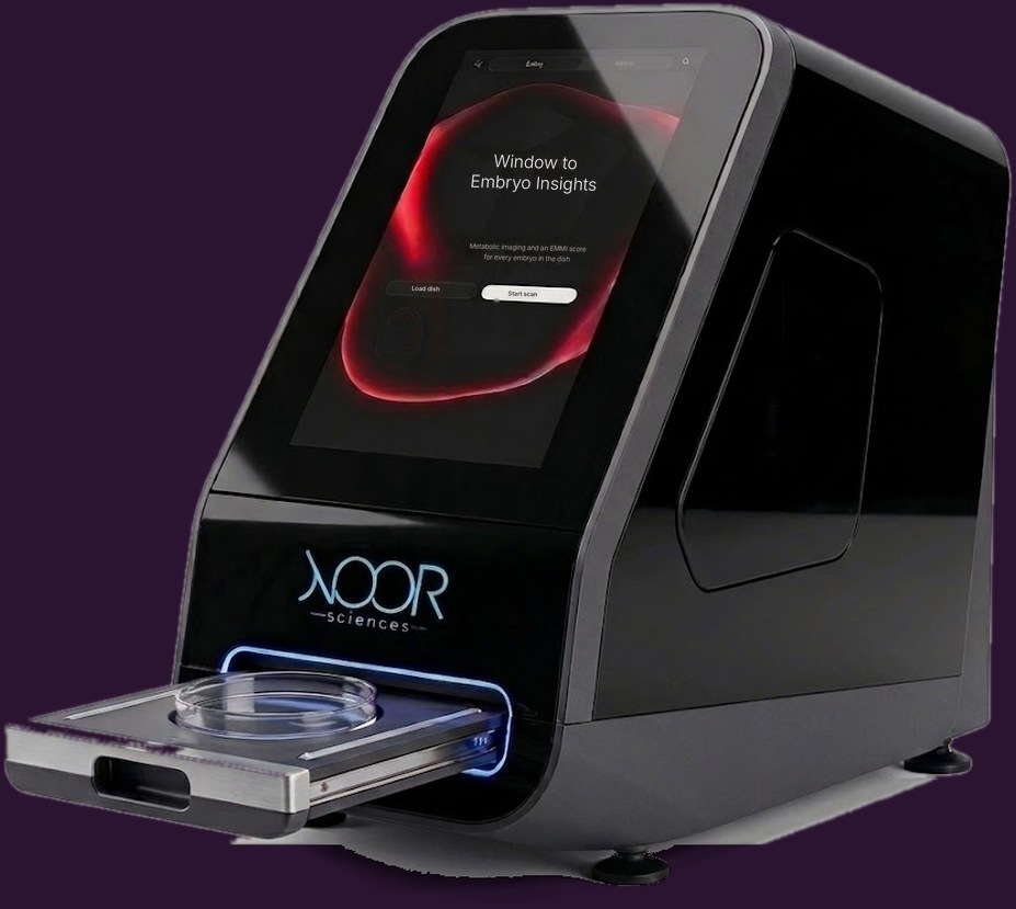
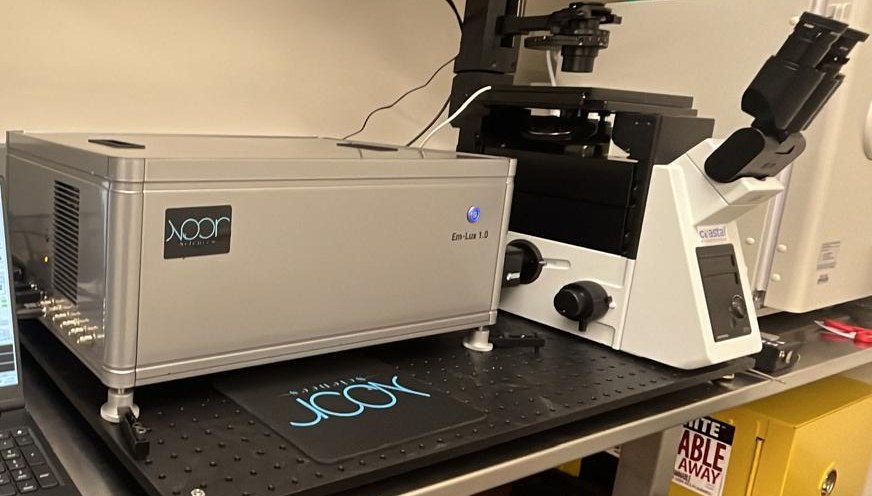
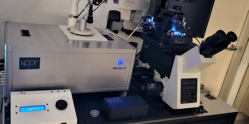
Em-Lux is a benchtop fluorescence lifetime imaging system. It images each embryo in its dish without dyes or contact, and EMMI, our algorithm, reports a score for each one. Not FDA cleared.
The instrument
Benchtop
A single benchtop unit for the embryology lab.
Completely non-invasive
Measures natural fluorescence. No dyes, stains or biopsy.
Imaging dish
Embryos are imaged in a single-use imaging dish.
Lifetime measurement
Fluorescence lifetime does not depend on signal brightness, which reduces sensitivity to optical setup.
From loading the dish to the report
- 01Set upSelect the patient and load the dish.
- 02LocateEm-Lux finds each embryo in the dish.
- 03AcquireBrightfield and metabolic images are captured for each one.
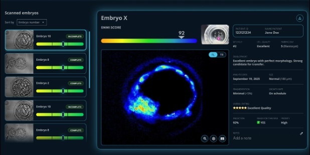
- 04ReportEach embryo receives an EMMI category (Low, Moderate or High) with its metabolic image. Embryos can be sorted by score, and the embryologist reviews.
EMMI is decision support. Final clinical decisions stay with qualified embryologists and physicians.
Embryo Morpho-Metabolic Index
EMMI is our proprietary algorithm. It is designed to weigh metabolic endpoints from Em-Lux together with morphology and clinical factors, assign each embryo a Low, Moderate or High category, and rank the cohort.
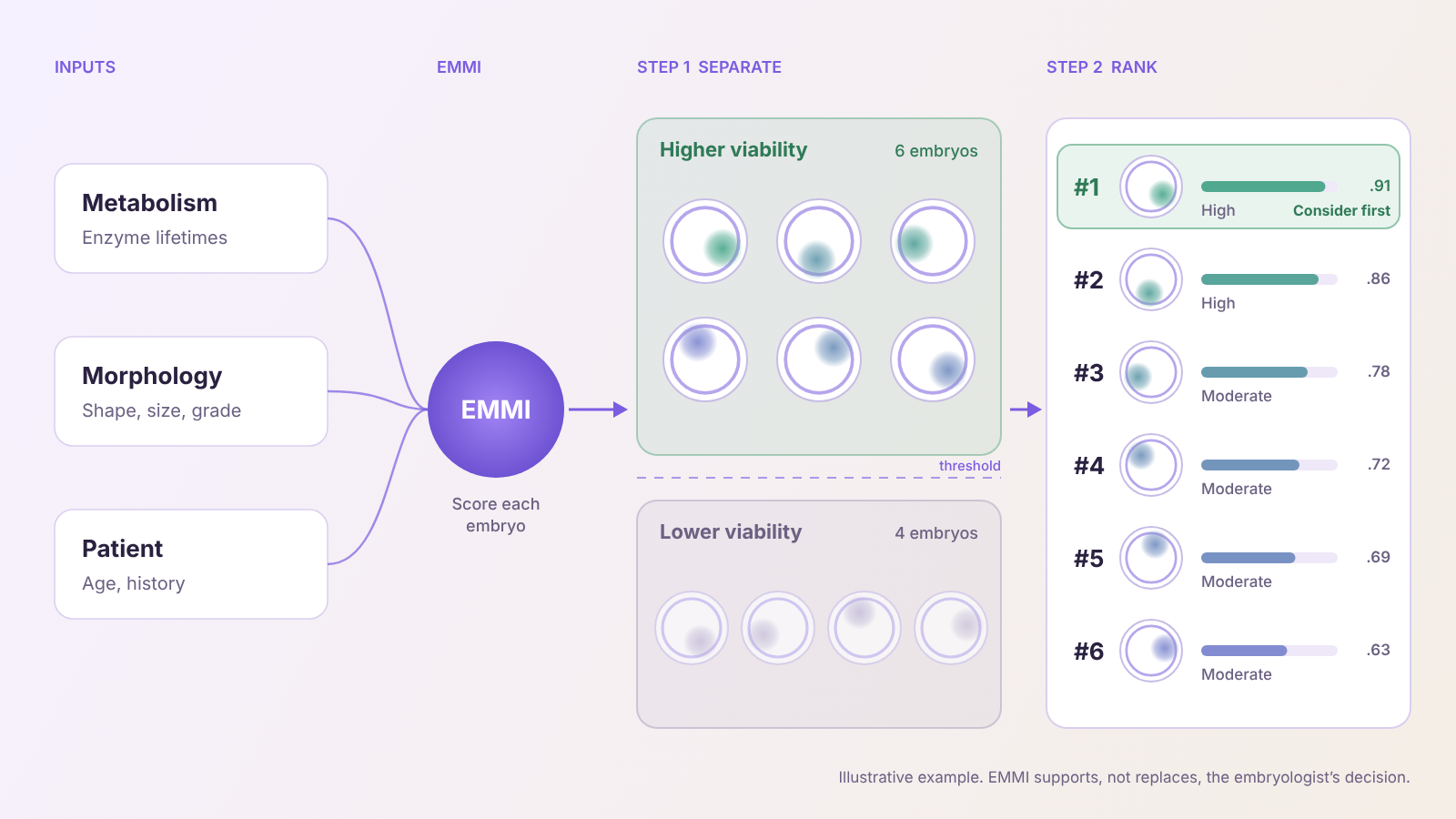
Inputs and outputs
How EMMI uses AI
Each scan goes through six steps, from image capture to a score with an explanation. The lifetime measurement is calibrated with physics, using no learned parameters, before the AI steps run. The embryologist makes the final decision.

Processing steps
- STEP 1AcquireLifetime and brightfield images
- STEP 2CheckAutomatic quality gate
- STEP 3CalibratePhysics layer, no learned parameters
- STEP 4LocateInner cell mass region
- STEP 5QuantifyMetabolic and morphologic features
- STEP 6ScoreEMMI score with explanation
FAQ
Is Em-Lux FDA cleared?
No. Em-Lux and EMMI are investigational and are not cleared or approved by the FDA.
Has the effect of imaging on embryos been studied?
Imaging safety has been studied in human embryos, and the results have been submitted for peer review. These are research data only. They have not been reviewed by the FDA.
Does it replace PGT-A?
No. PGT-A tests chromosomes and requires a biopsy. Em-Lux measures metabolism without contact. We expect them to be used together.
Can it assess eggs?
Egg assessment is in development with our academic partners and is not yet part of the product.
See where Em-Lux fits in your lab
Compare Em-Lux with grading, time-lapse and PGT-A, and see how clinics can take part in our clinical study.